


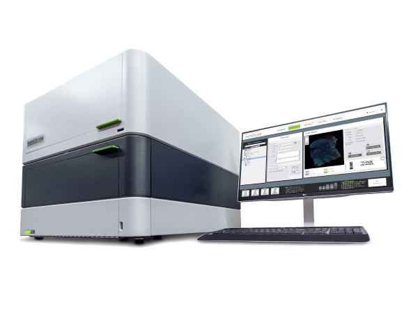


StrataQuest :
Context Tissue Cytometry
Context 유연한 조직 영상분석 소프트웨어로 종양미세환경을 넓게 탐색해보세요.
Feature
- 1
Import
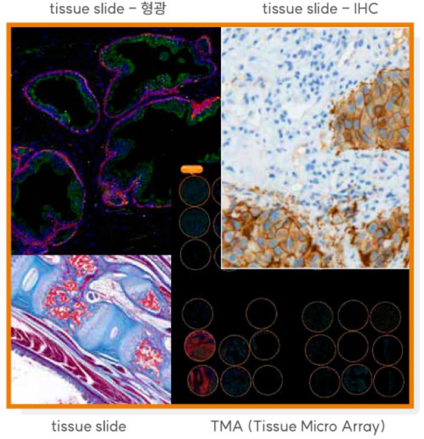
ㆍ 영상유형 : 명시야 및 형광영상
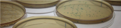
ㆍ 시료영상 : 조직슬라이드, TMA, 배양세포, Smear 등
- 2
유선형 single-cell 분석 소프트웨어 (IF & IHC App) : 조직영상에서 단일세포를 자동 구분 및 정량
ㆍ Deep-learning nuclear segmentation
ㆍ 영상 및 데이터 비교툴
ㆍ Image crops feature for scattergrams
ㆍ Z-stack and time-lapse analysis
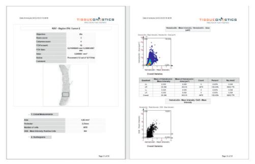
ㆍ 결과파일 : 보고서 pdf, 테이블 csv 및 이미지 tif, png 등 저장
- 3
머신러닝 조직분류기 및 meta-structure detection
- 4
맥락적 조직내 세포분석
ㆍ 거리 별 또는 근접 세포수 측정
ㆍ Tissue microenvironment 분석 및 공간적 분석
- 5
Spectral unmixing
- 6
Custom App : 원하는 형태로 분석절차를 구성하여 App으로 저장하여 자동실행
ㆍ 50여 가지의 표준분석 App
ㆍ 다양한 영상전처리 옵션
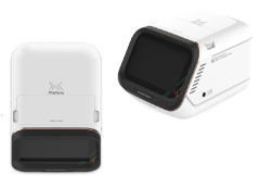
Import
다양한 포맷의 현미경 조직 이미지(IHC 및 형광)를 임포트 하여
맥락적 영상분석 (contextual image analysis)에 도전해 보세요.

- ㆍ TissueFAXS platform (*.aqproj)
- ㆍ StrataFAXS II (*.vmic)
- ㆍ PreciPoint (*.vmic, *.gtif)
- ㆍ Big TIFF (*.tif, *.tiff)
- ㆍ OME-Tiff (*.ome.tif, *.ome.tiff)
- ㆍ Image Folder (*.jpeg, *.png, *.bmp, *.tiff)
- ㆍ Zeiss (*.czi)
- ㆍ Hamamatsu NanoZoomer (*.ndpi, *.ndpis)
- ㆍ Leica Aperio (*.svs)
- ㆍ 3D HISTECH Pannoramic / Mirax (*.mrxs)
- ㆍ Olympus (*.vsi)
- ㆍ Perkin Elmer (*.qptiff, *.xml)
- ㆍ GE IN Cell Analyzer (*.xdce)
Single Cell Analysis
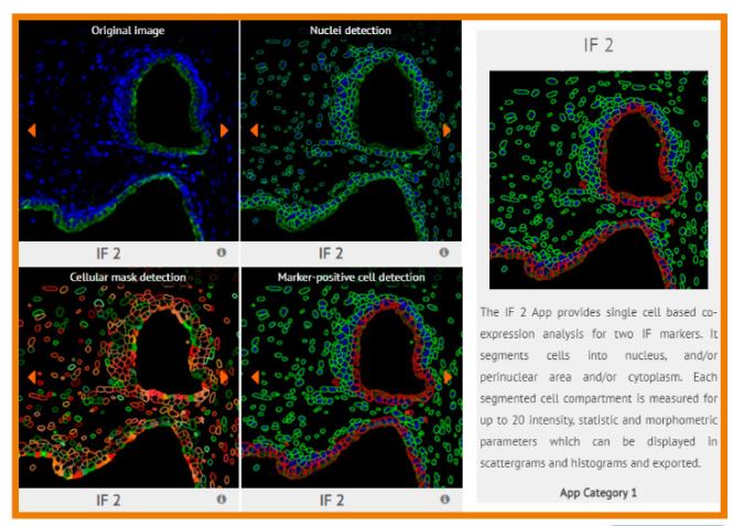
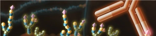
IHC이미지로부터 color separation 기법을 활용하여 색상을 분리하고 segmentation 기법으로 개별 세포의 핵 및 세포질을 분리한 후 스캐터 플롯 및 히스토그램으로 개별 세포특성을 통계 분석할 수 있습니다. 다중채널 형광이미지는 color separation 없이 segmentation 분석부터 시작할 수 있습니다.


조직슬라이드, TMA 및 배양세포용기
- Nuclear Segmentation
- Nuclear Segmentation Deep Learning
- Total Area Measurement
- Dot detection
- Membrane Detection

마커 또는 event에 따라 18개 기본 파라미터에서 지정하여 (기본 Average Intensity vs Area) 스캐터 플롯 및 히스토그램으로 표시합니다.
ㆍ Z stack 및 timelapse 데이터 분석
기본 파라미터
Area, Mean Intensity, Minimum of Intensity, Maximum of Intensity, Range of Intensity, Sum Intensity, Percentile, Percentile-Lower Mean, Percentile-Upper Mean, Variance of Intensity, STD of Intensity, Equivalent Diameter, Perimete, Compactness, Eccentricity, Minimum Width, Maximum Length, Feret Ratio
ㆍ Forward connection : 이미지상의 개별 세포를 클릭하면 스캐터 플롯상에서도 표시
ㆍ 영상 세트비교 툴

ㆍ 데이터 세트비교 툴

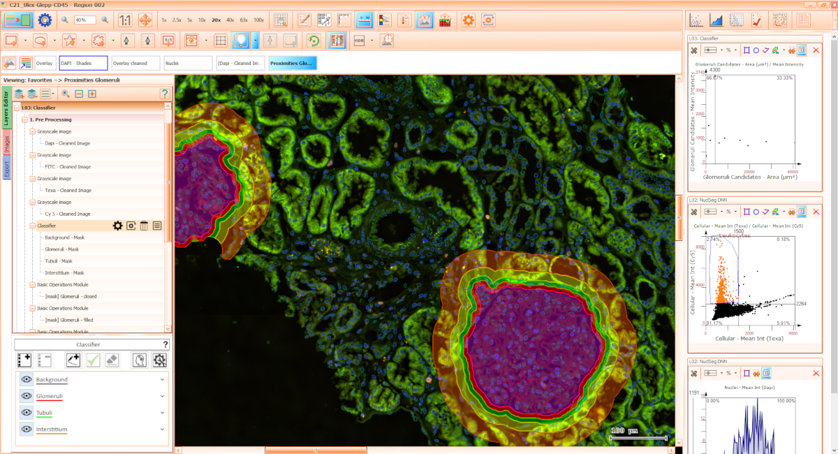
인공지능을 활용한 영상 세포학분석

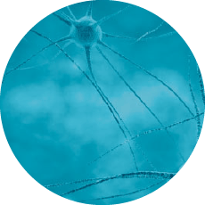
Deep Learning Nuclear Segmentation
세포가 작아 핵 구분이 쉽지 않은 조직 (특히 림프절 및 암조직) 또는 핵 내 염색질 분포가 불규칙한 경우 등 DNN (Deep Neural Network)를 활용하여 segmentation 할 수 있습니다.
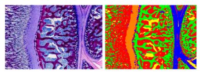
기계학습을 활용한 조직분류기
일부 조직영상에서 세포종류별로 지정하여 학습한 이후 전체 영상에 대해 자동으로 세포종류를 분류해줍니다. 학습된 프로파일은 다른 파일을 저장하고 불러올 수도 있습니다.
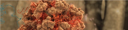
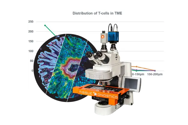
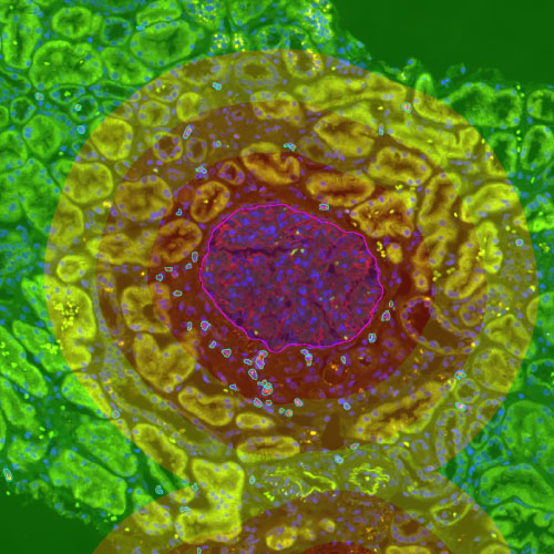
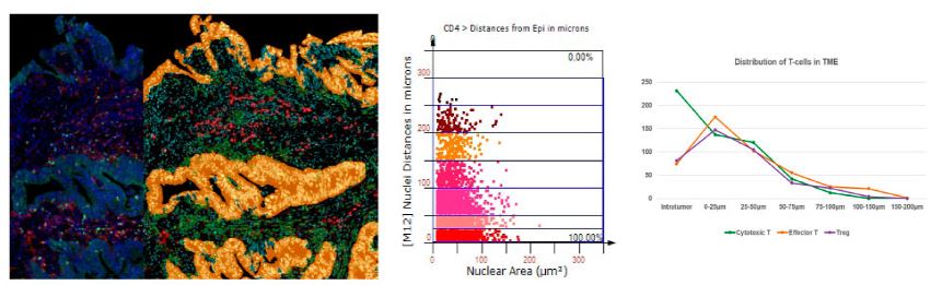
맥락적 조직내 세포분석
- Tumor Immune Microenvironment
-
ㆍ Distance mapping
암조직으로부터 거리 별로 면역세포의 분포를 체크하여 T cell infiltration 상황을 관찰하세요. -
PD-L1+ dendritic cells in the tumor microenvironment correlate with good prognosis and CD8+ T cell infiltration in colon cancer
Cancer Sci. 2020 Dec 20 참고문헌 보러가기 -
-
ㆍ Cell Connectivity Analysis
특정 면역세포로부터 일정거리 내에 있는 암세포의 수를 차트화하여 면역세포의 분포경향을 파악할 수 있습니다. 아래 예시는 triple negative vreast cancer 조직에서 CD20+ 세포가 50um 이내의 암세포수를 차트화 하였습니다. -
The spatial dynamics of the human tumor immune microenvironment (TIME)
EACR Congress, June 2021 참고문헌 보러가기 -

App
Custom App : 영상분석 및 정량절차를 App 파일로 저장하여 추후에 불러와서 사용할 수 있습니다.

표준 App : 다양한 기능의 App을 옵션으로 제공하여 필요 시 구매하여 분석할 수 있습니다.
APP 목록을 클릭하시면 아래 IF2 App처럼 내용을 살펴보실 수 있습니다.